One of the biggest challenges we face when it comes to motor neurone disease (MND) is the time it takes for people to be diagnosed.
At present, the length of time from the onset of symptoms to diagnosis is far too long – extending to months or even years - preventing early access to any available treatments and delaying people accessing the right support.
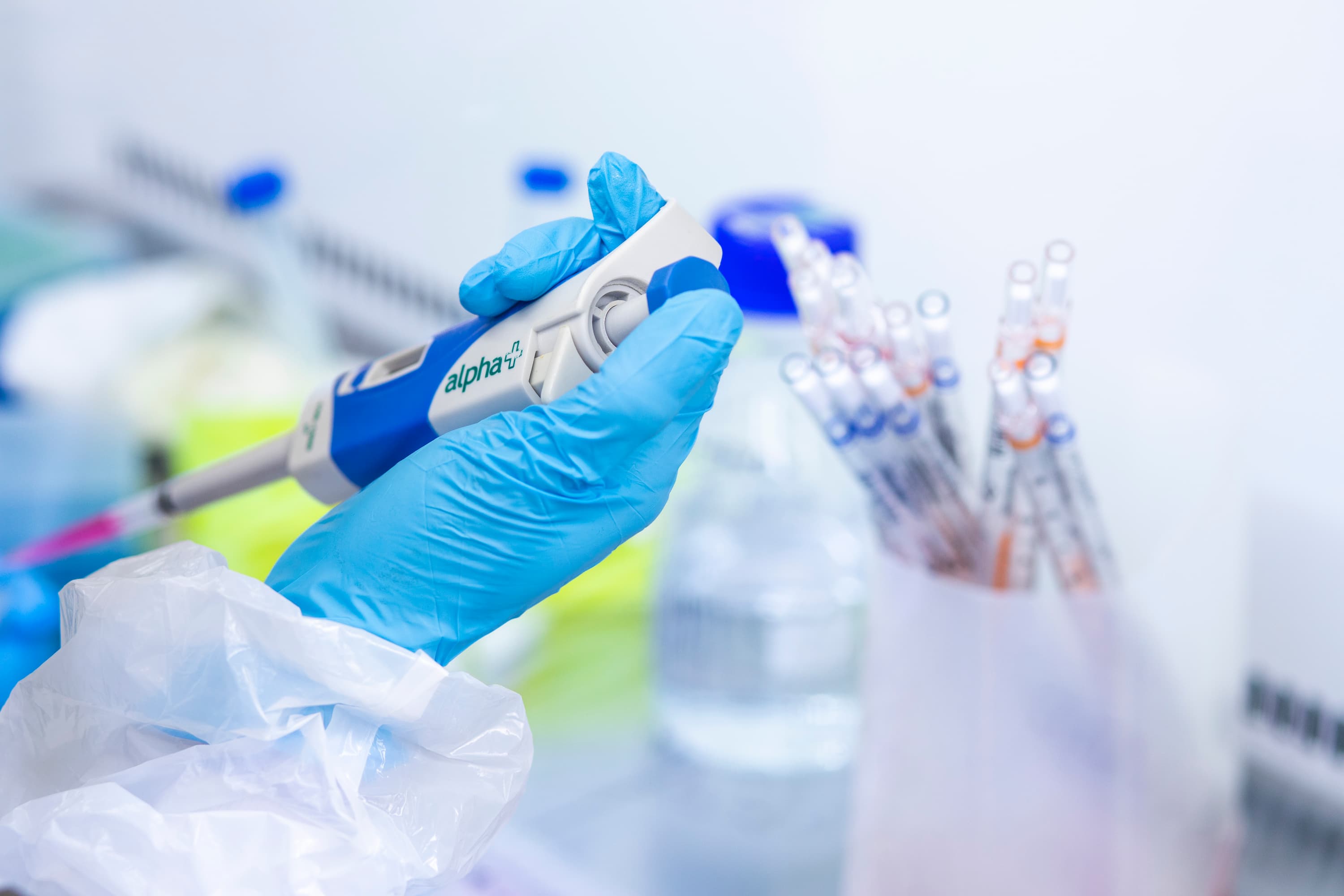
Among the many projects funded by the MND Association are ones investigating the use of imaging as a strategy for diagnosing MND. Using state-of-the-art technology, scientists are looking at ways to harness the power of scans to create detailed images of the brain and muscles to detect changes. Combined with a blood test in some cases to measure levels of neurofilament – a clear biomarker for nerve damage - these scans could hold the key to ensuring motor neurone disease is diagnosed at a much earlier stage in the future.

PhD student Dan Baxter-Beard and Principal Investigator, Professor Andrew Blamire at Newcastle University, whose work is being funded by the MND Association, are currently developing a new type of MRI scan which measures abnormalities in the muscles.
Using a Motor Unit MRI (MUMRI) the scan will identify the motor units within the limb which control the movement of muscles and identify any problems, usually within around three minutes. Crucially, the test is non-invasive and pain-free making it much easier for patients.

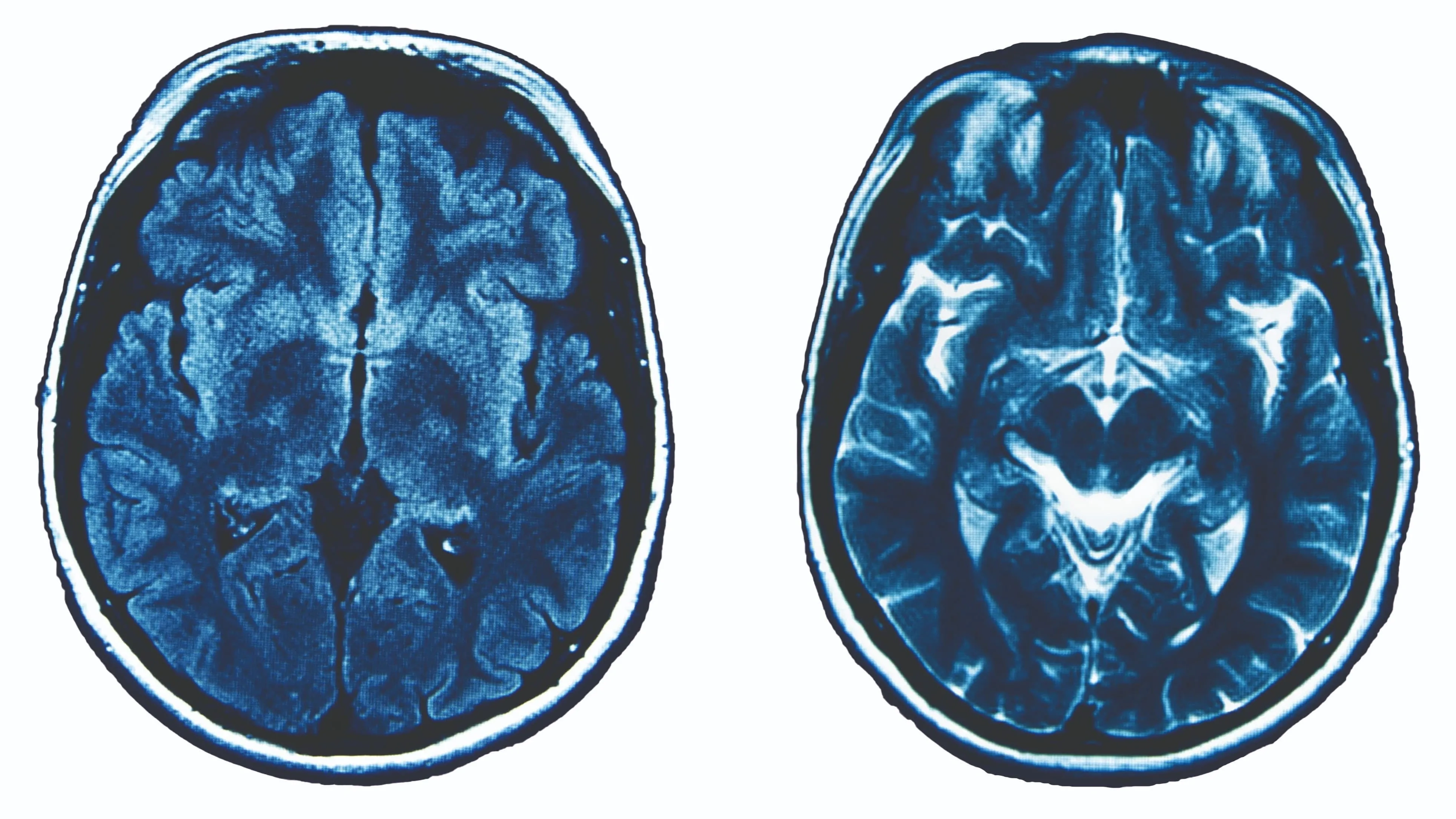
Another project, by PhD student Ayodeji Ijishakin and Principal Investigator Dr James Cole at University College London, is looking to use MRI scans to measure the effects of MND on brain volume and brain age. Combined with a blood test, it is hoped this method could be used to confirm the presence of MND, while also being used to measure the effectiveness of drug treatments if used as part of clinical trials in the future.

Meanwhile, Dr Marjorie Metzger at Trinity College Dublin is using a new brain imaging technique to see how the brain changes during the early stages of Primary Lateral Sclerosis (PLS) and MND. It is hoped that by using this new method to measure electrical activity in the brain, doctors will be able to distinguish between both conditions more easily, leading to earlier diagnosis and better ways to measure the effectiveness of potential treatments in clinical trials.
For more information about the progress we are making in this area visit our website.