We’ve all struggled to change a tyre or fix a tricky lightbulb and heard the expression: “it’s not rocket science!” But why not neuroscience? If you ask me, what goes on in the brain is far more mysterious than a rocket engine. After all, we made it to the moon but we’re still trying to make sense of the brain.
So, let me go back to basics, and explain a few of the trickier terms that you’ll have read in Prof Gareth Miles’ biography.
What is a neuron?
The simple explanation: a neuron is a nerve cell, found in the brain, that forms the basis of our nervous system. They transmit information and relay electrical signals, receive sensory input from the world around us and send motor commands to our muscles. At our best guess, our brains contain around 100 billion neurons.
Like other cells, they have a nucleus and a cell membrane, and they carry out basic cell processes such as energy production and protein synthesis.
Still with me? Great
Here’s where neurons differ. They are specialised cells and so contain parts that other cells don’t.
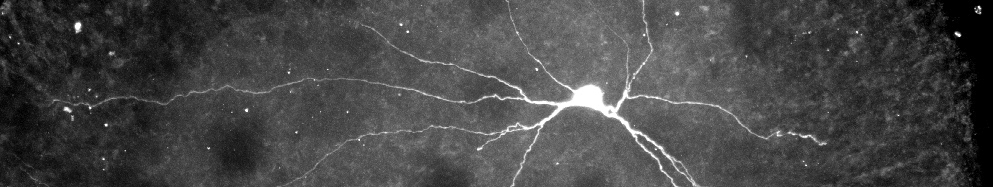
If you see a diagram of a common neuron, you’ll see shapes on one end that look like tree branches. These are known as ‘dendrites’ and that’s where the neurons receive most of their information, picking up signals from other neurons.
Those signals are then interpreted, and if the signal is strong enough it then travels through the cell and is transmitted by another part of the cell called ‘axons’.
In MND, neurons begin to degenerate and die, meaning they can’t send the signals that allow us to move our muscles. That’s why so much MND research is focussed simply on understanding how neurons work and process information.
Spinal circuits
Prof Gareth Miles and his team have been studying healthy neurons in the spinal cord in order to understand exactly what goes wrong in MND.
Healthy neurons are constantly transmitting and receiving information. In the spine, they come together to form spinal cord locomotor circuits. These transmit the signals needed to do all sorts of everyday activities, like walking and swimming.
But when the neurons degenerate, as they do in MND, the circuits and the connections between neurons break down. Prof Gareth Miles and his team want to understand why
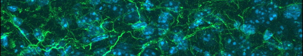
Glial Cells
Glial cells make up 90% of the cells in the brain. Scientists used to think they simply acted as a kind of insulation or glue holding the neurons together and allowing them to communicate. But we now know that glial cells are capable of communicating amongst themselves and sending information to neurons.
There are two types of glial cells, or ‘glia’. Microglia and macroglia. Microglia travel through the brain and spinal cord acting as an immune defence for the central nervous system. They remove things like pathogens (which cause disease) and damaged neurons.
There are also many types of macroglia. Here are a few:
Astrocytes – Astrocytes support the neurons and repair damage. They also help regulate communication between the neurons and help maintain a barrier between the brain and the blood.
Oligodendrocytes & Schwann cells – cells that make up the myelin sheath – a material that surrounds neurons to insulate electrical signals. Oligodendrocytes can be found in the central nervous system (the brain and spinal cord), and Schwann cells in the peripheral nervous system (nerves outside the central nervous system that link to organs, limbs and skin).
Researching glia is another way for scientists to understand how the brain and the nervous system function, and help us make sense of the process of degeneration that takes place in MND.